What Causes Osseous Demineralization? Symptoms and Treatment
Osseous demineralization happens when your bones gradually lose calcium and other minerals, often from aging, menopause-related estrogen loss, poor nutrition, inactivity, smoking, heavy alcohol use, chronic diseases, or long-term steroid/SSRI use. You might notice vague bone pain, fatigue, or weakness at first, then height loss, stooped posture, back pain, or fractures later. Doctors use DEXA scans and may prescribe medications plus lifestyle changes like exercise, calcium, and vitamin D—and you can find out exactly how those work next.
Key Takeaways
- Osseous demineralization is the loss of bone minerals like calcium and phosphorus, weakening bones and increasing fracture risk.
- Major causes include hormonal changes (especially post-menopause), poor calcium/vitamin D intake, inactivity, smoking, alcohol, chronic diseases, and certain medications.
- Early symptoms are subtle, such as vague bone pain, fatigue, and weakness; later signs include height loss, stooped posture, back pain, and fractures.
- Diagnosis relies mainly on central DEXA scans to measure bone mineral density, supplemented by X-rays and risk-factor assessment.
- Treatment combines medications (bisphosphonates, anabolic agents, denosumab, SERMs, hormone therapy) with lifestyle changes: adequate calcium/vitamin D, weight-bearing exercise, and avoiding smoking and heavy alcohol.
What Is Osseous Demineralization (And Why It Matters)
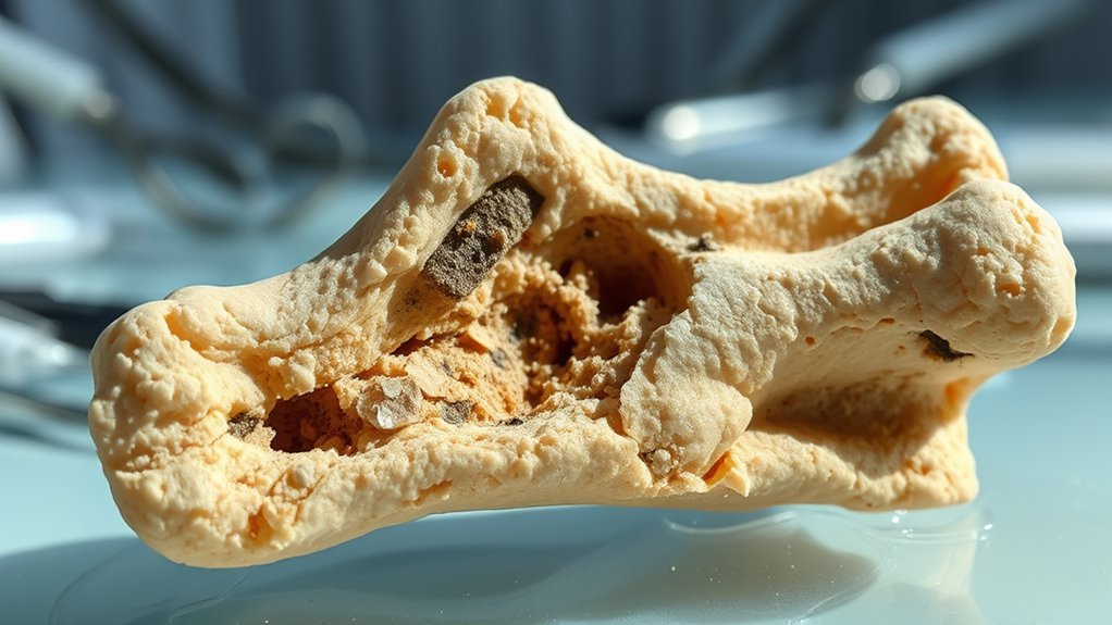
Osseous demineralization is the gradual loss of key minerals—mainly calcium and phosphorus—from your bones, and it quietly sets the stage for serious bone disease. Your skeleton normally stores about 99% of your body’s calcium and 85% of its phosphorus, constantly breaking down and rebuilding bone. When this balance is disrupted by factors such as aging, hormones, nutrition, or lifestyle, the risk of developing osteoporosis increases significantly.
Osseous demineralization slowly strips bones of calcium and phosphorus, silently paving the way for serious disease
When mineral loss outpaces replacement, bone mineral density (BMD) drops, your bones weaken, and fractures become more likely.
You’ll usually first see the impact as osteopenia, defined by a T-score between -1 and -2.5 on a central DEXA scan of your hip or spine. Osteopenia signals low BMD and higher fracture risk, but it’s still an early, more reversible stage.
If the demineralization continues, osteoporosis develops once your T-score falls below -2.5. At that point, fracture rates—especially hip and spine—rise sharply.
Detecting demineralization early with DEXA scanning helps you prevent this progression.
What Causes Osseous Demineralization in Adults?

Although bone loss can seem mysterious, osseous demineralization in adults usually traces back to a few well-defined drivers: shifting hormones, inadequate nutrition, lifestyle habits, chronic diseases, and certain medications or surgeries.
When estrogen drops after menopause or testosterone falls in men, bone breakdown speeds up. Too much thyroid or parathyroid hormone, or cortisol in Cushing syndrome, also pulls minerals from your skeleton. Early bone density assessment with tests like DEXA can help detect this mineral loss before it progresses to osteoporosis.
If you don’t get enough calcium, vitamin D, or protein—or you struggle with malnutrition or eating disorders—your body can’t maintain normal bone structure.
Daily choices matter too: inactivity, smoking, heavy alcohol use, extreme dieting, and prolonged bed rest all tip the balance toward bone loss.
Certain diseases directly undermine bone, including rheumatoid arthritis, chronic kidney disease, celiac disease and other gut disorders, diabetes, HIV/AIDS, and some cancers.
Long-term steroids, antiepileptics, PPIs, SSRIs, gastric bypass, and hormone-blocking cancer therapies further accelerate demineralization.
Who Is Most at Risk for Osseous Demineralization?
Knowing the main causes of bone loss naturally leads to another key question: who’s most likely to experience osseous demineralization in the first place?
You’re at highest risk if you were assigned female at birth, especially if you’re postmenopausal, over 50, or went through menopause early. Men aren’t exempt; low testosterone also speeds bone loss. Half of women over 50 will eventually develop osteoporosis, reflecting how common significant osseous demineralization is in this group.
Highest risk: individuals assigned female at birth after menopause—though men with low testosterone also lose bone faster
Age matters for everyone. Risk climbs after 50, and guidelines recommend routine screening for women 65+ and men 70+. Any fracture after 50 should trigger an evaluation.
Your build and background play a role. Caucasian and Asian women, people under about 125 pounds, those with a small frame, or low BMI face greater vulnerability.
Lifestyle and medical history compound risk: smoking, heavy alcohol use, low calcium or vitamin D, inactivity, eating disorders, family history of osteoporosis, chronic inflammatory or endocrine diseases, certain medications, and bariatric or major gastrointestinal surgery all greatly raise your odds.
Early and Late Symptoms You Shouldn’t Ignore
You mightn’t feel anything at first, but subtle early warning signs like vague bone discomfort, fatigue, or slight weakness can signal the beginning of osseous demineralization. Over time, progressive structural changes in your height and posture can reveal that your bones are losing strength. In many cases, these changes are linked to vitamin D deficiency, which reduces your body’s ability to absorb calcium and gradually weakens bone structure.
Subtle Early Warning Signs
How can a condition that quietly thins your bones announce itself before a major fracture happens? You may notice you’re a bit shorter—over time, your height drops more than 1.5 inches, clothes fit differently, or you appear smaller in photos.
A new stoop or hunched upper back, without an injury, can also signal weakening vertebrae.
Persistent back pain that flares when you bend, twist, or lift—especially without a clear cause—may reflect tiny compression fractures. Early diagnosis with a DEXA scan can detect low bone mineral density before symptoms become severe.
Pay attention if everyday mishaps cause outsized damage: a minor fall, a hard cough, or simple bending leads to a wrist, hip, or spine fracture.
Even reduced grip strength, like struggling to open jars or turn doorknobs, can quietly point to early bone loss.
Progressive Structural Changes
While early warning signs can be subtle, progressive structural changes speak much louder through your bones, muscles, and posture.
You may notice localized pain in your spine, hips, or legs that slowly worsens, or a deep ache in your lower back and pelvis. Anterior shoulder pain can sharpen with lifting or pushing, and your shins may feel unusually tender.
As muscle weakness progresses, climbing stairs, rising from a chair, or walking longer distances becomes harder.
Proximal leg weakness can cause a waddling gait and frequent stumbles.
Posture can shift into a stooped or forward‑bent position, often with measurable height loss.
Persistent fatigue, reduced endurance, and mobility limits that resemble arthritis or myopathy may further signal advancing demineralization.
Advanced Fracture-Related Symptoms
Even after the initial fracture pain settles, certain new or worsening symptoms can signal dangerous complications that need prompt attention.
Severe, deep pain that feels disproportionate to the visible injury, especially with tight swelling under a cast or splint, may indicate acute compartment syndrome—don’t wait if you notice increasing pain, numbness, or weakness.
Within 24–72 hours, sudden shortness of breath, confusion, or a new petechial rash can signal fat embolism syndrome.
Weeks to months later, persistent deep aching, inability to bear weight, or a visible bony bump may mean a nonhealing or malunited fracture.
Burning pain, color changes, and stiffness in a hand or foot suggest complex regional pain syndrome.
Ongoing fever, drainage, or escalating pain always warrants urgent evaluation.
How (And When) Osseous Demineralization Is Diagnosed
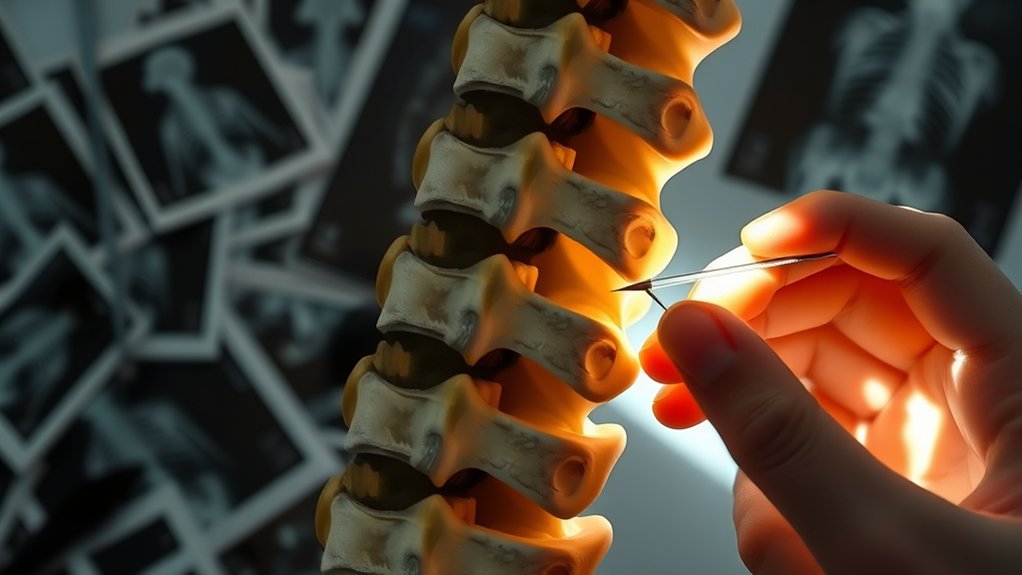
To understand how osseous demineralization is found early, you’ll start with key diagnostic tests, especially a central DEXA scan that measures bone density in your hip and spine.
Your doctor will recommend screening based on your age, medical history, medications, and any warning signs like fractures or height loss.
In many cases, you’ll get tested either because you meet routine screening criteria or because an incidental X-ray raises concern about bone loss.
Key Diagnostic Tests
Although osseous demineralization can affect many bones throughout the body, doctors rely on a few key tests and measurements to confirm it and gauge fracture risk. The gold standard is a central dual-energy X‑ray absorptiometry (DXA) scan, which measures calcium and other minerals in your spine and hip using low‑dose X‑rays. You’ll lie on a cushioned table while a scanner passes over you; it’s quick, painless, and usually finished within 15–30 minutes.
Peripheral devices can scan the forearm, wrist, fingers, or heel, but they’re less precise than central DXA. Results use T‑scores and Z‑scores, interpreted alongside your exam, medical history, and lab tests.
| Test/Measure | What It Evaluates | Key Points |
|---|---|---|
| Central DXA | Spine and hip BMD | Most accurate; gold standard |
| Peripheral DXA | Forearm, heel, wrist | Single site; less precise |
| T‑score | Density vs young adult peak | ≤ -2.5 indicates osteoporosis |
| Z‑score | Density vs age‑matched peers | Useful in younger patients |
| X‑ray | Existing fractures, bone shape | Can’t quantify mineral density |
When Screening Is Recommended
A DXA scan can tell you how strong your bones are, but knowing when to get that test matters just as much as how it’s performed.
Most guidelines say you should be screened at 65 if you’re a woman, or earlier if you’re postmenopausal with risk factors like parental hip fracture, low body weight, smoking, heavy alcohol use, certain medications, or conditions such as rheumatoid arthritis or intestinal disease.
If you’re 50–64 and need a chest X‑ray for another reason, your doctor may notice demineralization and suggest earlier DXA.
Medicare generally covers DXA every two years for women over 65 and men over 70. Your clinician will individualize how soon to start, and how often to repeat, based on your risk profile.
Medical Treatments to Stop or Reverse Osseous Demineralization
While lifestyle changes lay the foundation for bone health, medical treatments directly target the cells that build and break down bone to slow or even reverse osseous demineralization.
Bisphosphonates such as alendronate and risedronate (daily to monthly tablets), ibandronate (monthly tablet or quarterly IV), and zoledronic acid (yearly IV) slow bone breakdown and cut hip fractures by up to 40% and vertebral fractures by 30–70%.
If your bone loss is severe or you’ve had fractures, your doctor may suggest anabolic agents like teriparatide, abaloparatide, or romosozumab to actively build new bone for 1–2 years, then switch you to a bisphosphonate.
Denosumab, injected twice yearly, blocks formation of bone-dissolving cells but usually must continue indefinitely.
SERMs such as raloxifene or bazedoxifene, and in some women menopausal hormone therapy, reduce bone resorption and preserve density, especially after menopause.
Daily Habits to Protect and Prevent Bone Demineralization

Even without a prescription, the choices you make every day strongly influence how fast your bones lose or gain mineral.
First, hit your calcium targets: 1,000 mg daily for most adults and 1,200 mg for women over 50 and men over 70. Build meals around yogurt, cheese, almonds, dark leafy greens, or sardines with bones. Three cups of milk or fortified soy milk give you roughly 1,000 mg.
Pair that with 800–1,000 IU of vitamin D from fortified milk or cereal, fatty fish, eggs, and sunlight when appropriate to help you absorb calcium.
Move daily. Aim for 150 minutes a week of weight‑bearing exercise like brisk walking, stair climbing, dancing, or pickleball.
Protect your gains by limiting soda, excess caffeine, alcohol, salt, sugar, and processed foods, and by not smoking.
Emphasize protein, vitamin K2–rich natto, nettle tea, and low‑oxalate greens like kale and collards.
Frequently Asked Questions
Is Osseous Demineralization Reversible, and How Long Can Recovery Take?
It’s partly reversible, especially when you catch it early. You correct nutrition, exercise, and start medications; you’ll usually see improvements in 3–6 months, noticeable BMD gains in 6–24 months, then focus on long‑term maintenance.
Can Children or Teenagers Develop Osseous Demineralization, and Why?
Yes, you can develop osseous demineralization as a child or teen. It happens when poor nutrition, hormonal or endocrine disorders, medications, chronic illnesses, low activity, or eating disorders disrupt normal bone building during rapid growth.
How Does Osseous Demineralization Affect Dental and Jaw Health?
It weakens your jawbones, reduces bone density, and destabilizes teeth by accelerating alveolar bone loss. You’re more prone to fractures, shifting bite, periodontal disease progression, tooth loss, and you’ll often need more complex restorative or surgical dental treatments.
Are There Genetic Tests That Predict Future Bone Demineralization Risk?
You can’t currently take a direct genetic test that predicts future bone demineralization. Instead, you’d use DEXA scans, detailed family history, and sometimes broader osteoporosis‑risk genetic panels combined with clinical risk models.
Does Osseous Demineralization Increase Risks During Surgery or Anesthesia?
Yes, it does. You face higher fracture risk from fragile bones, anesthesia complications from electrolyte and hormonal imbalances, delayed emergence due to weakness and sarcopenia, and need tailored surgical planning with DEXA, medications, supplements, and careful mobilization.
Conclusion
Osseous demineralization isn’t just a medical term on your X‑ray report—it’s an early warning you shouldn’t ignore. By understanding the causes, watching for subtle symptoms, and getting timely testing, you can protect your bones before major damage occurs. Work closely with your healthcare provider, follow prescribed treatments, and build daily habits that support strong bones. When you take action early, you give yourself the best chance to maintain mobility, independence, and long‑term health.